

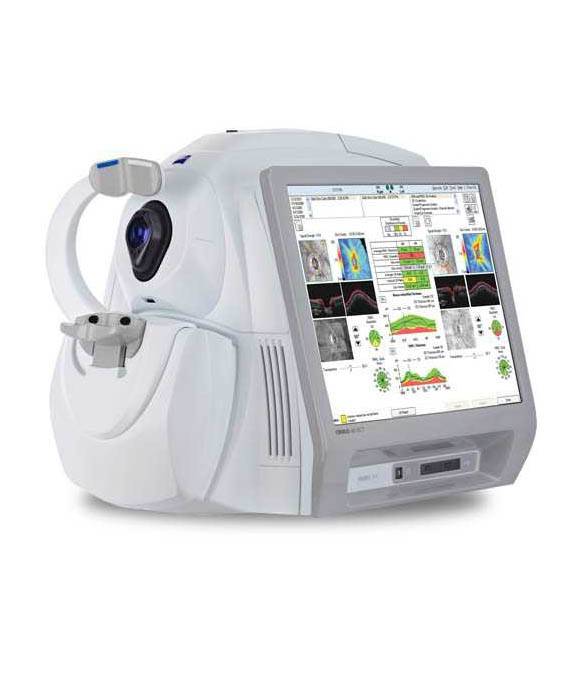
OPTICAL COHORENS TOMOGRAPHY
Optical Coherence Tomography, otherwise known as OCT, is an advanced crosssectional imaging method used to examine and measure intraocular structures.
Although the working principle of Optical Coherence Tomography (OCT) is similar to the ultrasound applications applied to the eye, unlike the sound waves used in ultrasound, spectrofotmetric light of 820 nanometer wavelength is used in OCT.
Because the speed of light is about 1 million times faster than the speed of sound, the OCT's lower resolution of 10 nanometers provides more efficient results than the 100 nanometer resolution of ultrasound.
Unlike ultrasonography, OCT can be applied to the non-dilated pupil and there is no need for eye drops and contact to the eye.
MEASUREMENT WITH OPTICAL COHERENS TOMOGRAPHY
In Optical Coherence Tomography, the delay times of the reflection of the light emanating from the light center are measured. In particular, it allows detailed analysis of the optic nerve, retina and macula. Optical Coherence Tomography is widely used for the diagnosis of age-related macular degeneration due to systemic diseases and the condition of the disease. If you wish, you can take a look at the age-related macular degeneration (AMD) article published in a different title on our website.
For OCT devices with a longer wavelength light center for the front of the eye. This advanced method can provide higher resolution images and measurements of cornea, iris and intraocular instruments and lenses.