COLOR BLINDNESS
COLOR BLINDNESS TREATMENT
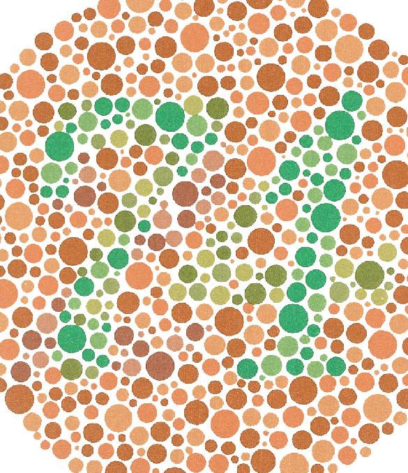
Color blindness, otherwise called Daltonism, occurs in cone photoreceptors in the retina. Color blindness test process consists of colored plates, aka Ishihara plates, which contain a number among dots, randomized in size and color. Color blindness can be diagnosed if individuals cannot read the numbers and figures on the plates. This test is the most widely used method in the diagnosis of color blindness.
The test in which individuals read plates composed of numbers and objects written in different colors is called a color blindness test. If individuals are unable to read the numbers and shapes on the plates, they may be diagnosed with color blindness. The most commonly used method for diagnosing color blindness is the color blindness test conducted with physical displays, as mentioned above.
Causes of Color Blindness
Color blindness, which is estimated to be found in approximately 170-180 million people around the world, is a disorder characterized by the absence or scarcity of pigment particles that should be in the visual center of the brain. The individuals with color blindness may not be able to distinguish one or more colors. Color blindness is a genetic condition.
Types of Color Blindness
Color blindness can be divided in different groups;
- “Protonopia" : red color blindness due to red cone photoreceptors,
- “Deuteranopia” : red color blindness due to green cone photoreceptors,
- “Tritonopia" : red color blindness due to blue cone photoreceptors,

These color blindness types indicate partial color blindness. However, in cases of monochromatic color blindness, complete color blindness can be mentioned, and when people are completely color blind, they cannot distinguish colors from each other under any circumstances (except for black and white).

